Recent publications, including the 9th edition, emphasize the crucial balance between diagnostic benefits and minimizing radiation exposure.
Essential physics and radiation protection are thoroughly covered, guiding professionals in safe imaging practices, as of January 24, 2026.
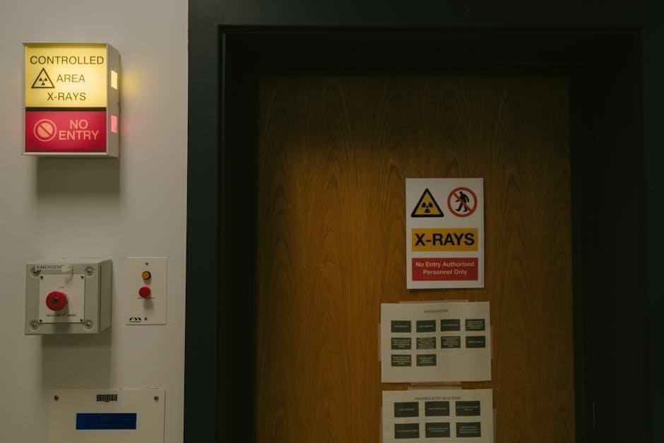
Historical Context of Radiation Safety
Early radiography, pioneered in the late 19th century, lacked robust safety protocols, leading to significant health consequences for both patients and practitioners. Initial awareness stemmed from observed tissue damage – burns and ulcerations – prompting initial, albeit rudimentary, protective measures.
The field evolved through decades of research into the biological effects of ionizing radiation, culminating in the establishment of international guidelines and national regulations. Publications, like the evolving editions of “Radiation Protection in Medical Radiography”, reflect this progression.
The 9th edition builds upon this history, acknowledging past lessons and integrating contemporary advancements in technology and understanding of radiation-induced non-cancer diseases, emphasizing a continuous commitment to safety.
The Importance of the 9th Edition
The 9th edition of “Radiation Protection in Medical Radiography” is paramount, reflecting the latest advancements in imaging technology and a deepened understanding of radiation’s biological impacts. It addresses evolving regulatory landscapes and emphasizes optimized patient safety protocols.
This edition consolidates knowledge regarding stochastic and deterministic effects, alongside insights into radiation-induced non-cancer diseases. It’s crucial for professionals navigating the complexities of dose management, particularly in paediatric imaging, where sensitivity is heightened.
Staying current with this resource ensures adherence to national and international guidelines, fostering responsible practice and minimizing unnecessary radiation exposure.

Fundamental Principles of Radiation Protection
Core tenets include justification of practices, optimization via ALARA, and strict adherence to established dose limits – foundational for safe radiography.
Justification of Practices
The principle of justification demands that any medical exposure to radiation must deliver a demonstrable benefit to the patient, outweighing the potential risks. This isn’t simply about performing an exam; it requires careful consideration of the clinical indication and whether alternative, non-ionizing imaging modalities could provide equivalent diagnostic information.
Recent publications, including updated editions, reinforce this concept, emphasizing the need for a documented rationale for each radiographic procedure.
Radiographers must actively participate in this process, questioning requests that lack clear clinical justification and advocating for the most appropriate imaging strategy for each individual patient, ensuring responsible radiation use.
Optimization of Protection (ALARA)
ALARA – As Low As Reasonably Achievable – is the cornerstone of radiation protection, demanding continuous efforts to minimize dose without compromising image quality. This involves meticulous technique selection, utilizing shielding effectively, and employing time and distance principles.
The 9th edition of relevant texts highlights advancements in digital radiography and AI, offering opportunities for significant dose reduction.
Radiographers are responsible for implementing ALARA through collimation, filtration, and careful patient positioning, always striving to deliver the lowest possible radiation dose for a diagnostically acceptable image.
Dose Limits and Regulations
National and international guidelines establish strict dose limits for both occupational exposure and the public, ensuring radiation safety. These regulations, frequently updated in publications like the 9th edition resources, are crucial for protecting patients and personnel.
Compliance requires diligent monitoring of radiation levels, adherence to prescribed protocols, and comprehensive quality assurance programs.
Radiation Safety Officers play a vital role in enforcing these standards, maintaining records, and providing ongoing training to radiography staff, guaranteeing responsible radiation practices.
Biological Effects of Radiation
The 9th edition details both stochastic effects – like cancer risk – and deterministic effects, or tissue reactions, following radiation exposure.
Non-cancer diseases are also modeled within a radiation protection context.
Stochastic Effects (Cancer Risk)
Stochastic effects, primarily cancer risk, are probabilistic, meaning their likelihood increases with dose, but severity isn’t dose-dependent. The 9th edition thoroughly examines radiation cancerogenesis incidence, providing crucial data for risk assessment in medical radiography.
Understanding these effects is paramount for justifying imaging procedures and optimizing patient protection. Publications emphasize the importance of minimizing exposure, even at low doses, due to the potential for long-term cancer development.
The edition likely details factors influencing individual susceptibility and provides updated models for estimating cancer risk based on radiation dose, aligning with current regulatory guidelines and best practices.
Deterministic Effects (Tissue Reactions)
Deterministic effects, or tissue reactions, have a dose threshold; below this, the effect doesn’t occur, and above it, severity increases with dose. The 9th edition likely details these effects, such as skin erythema and cataracts, relevant to radiography.
Understanding thresholds for various organs is crucial for preventing these predictable, non-stochastic outcomes.
The text probably emphasizes techniques to avoid exceeding these thresholds through careful technique selection, shielding, and patient positioning.
Modern radiography aims to eliminate deterministic effects entirely through adherence to established dose limits and rigorous quality assurance programs.
Radiation-Induced Non-Cancer Diseases
The 9th edition of resources on radiation protection likely addresses radiation-induced non-cancer diseases, acknowledging potential long-term health risks beyond malignancy. These effects, while less common than stochastic risks, are important considerations.
Publications detail models examining incidence following radiation exposure, potentially including cardiovascular disease and cataracts.
Understanding these risks informs strategies for minimizing overall patient exposure, even at low doses.
The text likely emphasizes the importance of optimizing protocols and employing ALARA principles to mitigate the possibility of these delayed health consequences.
Radiation Protection Techniques in Radiography
Effective techniques, such as shielding, distance, and time minimization, are central to safe radiography practices, as detailed in current publications.
Shielding Principles (Lead Equivalency)
Shielding is a cornerstone of radiation protection, utilizing materials like lead to attenuate X-ray beams and safeguard personnel and patients. The concept of lead equivalency is vital; it defines the thickness of another material needed to provide the same degree of attenuation as a specific thickness of lead.
Understanding lead equivalency allows for the use of alternative shielding materials, potentially lighter and more cost-effective, while maintaining equivalent protection levels. Current resources, including the 9th edition materials, emphasize proper shielding design based on factors like X-ray tube output, distance, and workload, ensuring compliance with regulatory standards and minimizing unnecessary exposure.
Distance and Inverse Square Law
Distance is a primary factor in radiation protection, governed by the inverse square law. This principle dictates that radiation intensity decreases dramatically as the distance from the source increases – specifically, inversely proportional to the square of the distance. Doubling the distance reduces the intensity to one-fourth.
The 9th edition resources highlight maximizing distance as a simple yet highly effective method for reducing exposure. Radiographers should utilize available distance whenever possible during exposures, and understand its impact on radiation dose. Proper positioning and utilizing control panels outside the primary beam are key applications of this fundamental principle.
Time Minimization
Time is a critical component of radiation protection, directly influencing cumulative dose. Minimizing exposure time reduces the total amount of radiation received by both patient and personnel. The 9th edition materials emphasize efficient workflow and technique optimization to achieve this.
Reducing exposure time doesn’t necessarily mean rushing; it means preparedness. Proper patient positioning, accurate technique selection, and minimizing fluoroscopic time are essential. Utilizing techniques like pulsed fluoroscopy further reduces dose. Prioritizing efficient image acquisition is paramount for upholding ALARA principles.
Equipment and Radiation Protection
Modern radiography equipment features enhanced filtration, collimation, and image receptor technology, all contributing to significant dose reduction, as detailed in recent editions.
X-ray Tube Design and Filtration
X-ray tube design significantly impacts radiation output and quality, directly influencing patient dose and image formation. Modern tubes incorporate improved anode materials and focal spot configurations for enhanced efficiency. Filtration, a critical component, selectively removes low-energy photons that contribute to patient dose without adding diagnostic information.
Aluminum filtration is standard, with options for added filtration based on imaging parameters and patient size. The 9th edition resources emphasize inherent filtration within the tube housing, alongside added filtration, to optimize beam quality; Proper filtration minimizes skin dose and improves image contrast, aligning with ALARA principles.
Understanding these design elements is fundamental for radiographers to implement effective radiation protection strategies.
Collimation and Beam Limitation
Collimation is a cornerstone of radiation protection, restricting the x-ray beam to the area of clinical interest. Positive Beam Limitation (PBL) is a mandatory feature on modern radiographic equipment, ensuring the beam cannot be larger than the image receptor. Accurate collimation directly reduces patient dose by minimizing scatter radiation and exposing only the necessary anatomy.
The 9th edition materials highlight the importance of utilizing collimation effectively, alongside other techniques, to adhere to ALARA principles. Proper beam limitation not only protects patients but also enhances image quality by reducing scatter, improving contrast, and reducing artifacts.
Radiographers must prioritize precise collimation in every examination.
Image Receptor Considerations
Image receptor technology significantly impacts radiation dose. Digital radiography (DR) and computed radiography (CR) systems generally require lower radiation doses compared to traditional film-screen systems to achieve equivalent image quality. The 9th edition emphasizes understanding the inherent dose characteristics of each receptor type.
Detector efficiency, the ability to convert x-ray photons into a signal, is a key factor. Higher efficiency receptors require less radiation. Furthermore, proper receptor selection and utilization, alongside collimation and filtration, contribute to optimized patient protection.
Radiographers must be knowledgeable about receptor limitations.
Patient Dose Management
Effective dose calculations and Dose Area Product (DAP) measurements are vital for assessing and managing patient radiation exposure, as detailed in recent editions.
Dose Area Product (DAP)
Dose Area Product (DAP) is a crucial metric in medical radiography for quantifying the total radiation delivered to a patient during a fluoroscopic procedure. It represents the integrated dose over the irradiated area, expressed in Gy·cm2.
Understanding DAP values allows for objective assessment of radiation exposure, facilitating comparisons between different examinations and patients. Monitoring DAP helps ensure adherence to established dose reference levels and supports optimization of radiation protection practices.
Recent publications, including updated editions, emphasize the importance of DAP monitoring as a key component of comprehensive patient dose management programs within radiology departments. Accurate DAP readings contribute to safer imaging protocols.
Effective Dose Calculations
Effective dose estimations are paramount in medical radiography, representing the risk of stochastic effects – primarily cancer – from radiation exposure. Calculated using tissue weighting factors, it accounts for varying sensitivities of different organs and tissues to radiation.
The 9th edition resources highlight the importance of accurate effective dose calculations for optimizing patient protection. These calculations move beyond absorbed dose to provide a more clinically relevant measure of potential harm;
Understanding effective dose enables informed risk-benefit analyses, particularly in paediatric imaging, ensuring justifiable practices and minimized patient risk, as emphasized in current guidelines.
Pediatric Radiation Protection
Pediatric patients are significantly more radiosensitive than adults, demanding heightened attention to radiation protection principles. The 9th edition resources strongly advocate for minimizing exposure in children due to their longer life expectancy and developing tissues.
Risk-benefit analysis is crucial when considering paediatric imaging, carefully weighing the diagnostic value against potential radiation risks. Techniques like collimation, shielding, and lower technique settings are essential.
Effective communication with parents, providing informed consent, and addressing concerns are vital components of responsible paediatric radiography, as detailed in oral radiology principles.

Regulatory Framework and Standards
National and international guidelines dictate radiation safety protocols, while robust quality assurance programs ensure compliance.
Radiation Safety Officers bear responsibility for upholding these standards, as outlined in current publications.
National and International Guidelines
Globally, radiation protection standards are built upon recommendations from international bodies, striving for consistent safety measures across borders. These guidelines, frequently updated and reflected in the 9th edition resources, emphasize justification and optimization principles.
National regulations, such as those enforced by governing health agencies, translate these international recommendations into legally binding requirements for medical radiography facilities. These regulations cover aspects like equipment calibration, personnel training, and dose monitoring.
Compliance with these standards is paramount, ensuring patient and staff safety while maintaining the diagnostic quality of imaging procedures. Ongoing professional development and adherence to published guidelines are essential for responsible practice.
Quality Assurance Programs
Robust Quality Assurance (QA) programs are fundamental to maintaining optimal radiation safety and image quality in medical radiography, as detailed in current editions. These programs involve systematic monitoring and evaluation of all aspects of the imaging process.
Regular testing of X-ray equipment, including tube output, collimation accuracy, and filtration effectiveness, is crucial. QA also encompasses meticulous record-keeping of doses, incidents, and corrective actions taken.
Effective QA requires a dedicated team and a commitment to continuous improvement, ensuring adherence to national and international guidelines. These programs directly contribute to minimizing patient exposure and maximizing diagnostic information.
Radiation Safety Officer Responsibilities
The Radiation Safety Officer (RSO) holds a pivotal role in ensuring compliance with all applicable radiation safety regulations, as outlined in the latest radiography resources. Their duties encompass overseeing the facility’s radiation protection program, including personnel monitoring and dose tracking.
RSOs are responsible for conducting regular safety audits, investigating incidents involving radiation exposure, and implementing corrective actions. They also provide comprehensive training to all staff on proper radiation safety procedures.
Maintaining accurate records, staying current with regulatory changes, and serving as a liaison with regulatory agencies are also key RSO responsibilities.
Communicating Radiation Risks
Effective communication, including patient education and informed consent, is vital, particularly in paediatric imaging, to address concerns and explain risk-benefit analyses.
Patient Education and Informed Consent
Comprehensive patient education is paramount in modern radiography, ensuring individuals understand the rationale behind imaging procedures and associated radiation exposure. The 9th edition likely reinforces the necessity of transparent communication regarding potential risks, however minimal.
Informed consent transcends simply obtaining a signature; it demands a clear, accessible explanation of the benefits versus risks, tailored to the patient’s understanding. This includes discussing alternative imaging modalities, if available, and addressing any anxieties or misconceptions.
Effective communication builds trust and empowers patients to actively participate in their healthcare decisions, fostering a collaborative approach to radiation safety and responsible imaging practices.
Risk-Benefit Analysis in Paediatric Imaging
Paediatric imaging necessitates a particularly rigorous risk-benefit analysis due to increased sensitivity to radiation. The 9th edition likely emphasizes minimizing exposure while obtaining diagnostically valuable images. Careful consideration must be given to the clinical indication, justifying the examination and selecting the lowest possible dose.
Alternative modalities, such as ultrasound or MRI, should be explored whenever feasible to avoid ionizing radiation altogether. When radiography is essential, techniques like collimation, shielding, and optimized protocols are crucial.
Thorough documentation of the justification and dose optimization strategies is vital, demonstrating a commitment to paediatric radiation protection principles.
Addressing Patient Concerns
Effective communication is paramount when addressing patient anxieties regarding radiation exposure; The 9th edition likely reinforces the importance of clear, empathetic explanations about the benefits outweighing the risks. Patients deserve honest information presented in an understandable manner, avoiding technical jargon.
Acknowledging concerns and actively listening to patient questions builds trust. Radiographers should be prepared to discuss dose reduction strategies and the safety measures in place.
Providing reassurance and emphasizing the skilled expertise of the imaging team can alleviate fears, fostering a positive patient experience.
Future Trends in Radiation Protection
Digital radiography and artificial intelligence are poised to optimize dose levels, alongside advancements in shielding materials, enhancing patient safety continually.
Digital Radiography and Dose Reduction
Digital radiography represents a significant leap forward in radiation protection, offering inherent dose reduction capabilities compared to conventional film-screen systems. This stems from the increased detective quantum efficiency (DQE) of digital detectors, requiring less radiation to produce an image of comparable quality.
Furthermore, post-processing techniques allow for image manipulation and optimization, potentially reducing the need for repeat exposures. The 9th edition likely details these advancements, emphasizing the importance of utilizing digital technology’s full potential.
Ongoing research focuses on further refining digital systems and algorithms to minimize patient dose while maintaining diagnostic accuracy, aligning with the ALARA principle.
Artificial Intelligence in Dose Optimization
Artificial intelligence (AI) is rapidly emerging as a powerful tool in radiation protection, particularly in optimizing patient dose during medical radiography. AI algorithms can analyze image data and automatically adjust exposure parameters to deliver the lowest possible dose while maintaining diagnostic image quality.
These systems can learn from vast datasets of images and clinical information, identifying patterns and predicting optimal settings for individual patients. The 9th edition likely explores these applications, highlighting AI’s potential to personalize dose levels.
Further development promises even more sophisticated AI-driven dose management strategies, enhancing patient safety and aligning with ALARA principles.
Advancements in Shielding Materials
Ongoing research focuses on developing novel shielding materials that offer improved radiation attenuation with reduced weight and thickness compared to traditional lead. These advancements are crucial for enhancing protection for both patients and radiographers.
The 9th edition may detail explorations into alternative materials like tungsten-based alloys, bismuth, and polymer composites incorporating high-density elements. These materials aim to provide equivalent protection while addressing concerns about lead’s environmental impact and weight.
Improved shielding contributes to a safer working environment and reduced scatter radiation, ultimately optimizing radiation protection protocols.